")
The technology used in modern dentistry is always changing and progressing. Many dentists stay on top of these technological advances, using them to create better patient experiences and outcomes.
This month, we take a look at some of the newer 3D imaging technologies, including cone beam technology, that are making dental surgeries easier.
Why 3D Imaging in Dentistry?
As with most medical imaging, 3D imaging takes a series of pictures or snapshots and then uses computers to piece together a 3D model or image. 3D imaging gives dentists a much better view of the mouth and jaw, allowing them to better see and diagnose problems, and to determine the next step in treatment.
One of the most exciting kinds of 3D dental imaging these days is called “Cone Beam Computed Tomography” or CBCT.
What is Cone Beam Computed Tomography (CBCT)?
Cone beam computed tomography is a 3D medical imaging technique that uses traditional X-ray computed tomography but spreads the X-rays out in a cone shape (hence the name). This allows the dentist to get a more complete model of a patient’s face, jaw, and teeth.
Think of it as being similar to a smartphone that takes a panoramic picture. The phone actually takes a series of smaller pictures as you move the phone, and then stitches the smaller images together into one panoramic shot. In a similar way, CBCT takes several pictures of a patient’s mouth and jaw by rotating around the patient’s head. But, with CBCT, those pictures are taken using X-rays to see the underlying teeth and bone structure. Computers are then used to stitch together the complete image.
CBCT has become increasingly important in diagnosis and treatment planning, particularly for implants, surgery, and orthodontics.
“I cannot imagine practicing without a cone beam machine in my office,” says Michael Simon, DMD, with Prosthodontic Associates of South Florida. “Every patient that I have scheduled to perform implant surgery has a cone beam [CBCT] image done for their specific surgical site. I would go as far as to say that I will not perform implant surgery on a patient without a cone beam radiograph.” Simon mentions that he uses a Planmeca digital panorex machine in his practice.
Simon also says he routinely uses CBCT technology for patients with pain symptoms that cannot be easily diagnosed using more traditional 2D radiographs (apical radiolucencies and fractures, for example, that do not readily appear on a two-dimensional film).
What is a CBCT Scanning Session Like for the Patient?
Today’s medical imaging can seem otherworldly to most patients. But the process is incredibly safe, comfortable, and accurate. Here’s what an imaging patient can reasonably expect with the process:
Getting comfortable in the machine. Depending on the type of cone beam imaging system used, a patient might be standing or sitting as the machine’s “camera” is put into position. The patient’s head is stabilized with a chin rest (or something similar), to minimize movement during the scan and to keep the patient comfortable.
Performing the scan. The X-ray “camera” rotates around the patient’s head on one side while a detector on the opposite side rotates in tandem with it. This is when a patient must remain as still as possible. Note that, during scanning, there might be some noises associated with the working of the machine. That said, the scan itself usually takes seconds. Radiation exposure is minimal, and much less than traditional X-rays.
Data processing and image building. The detector picks up the X-ray images generated—up to 600 of them!—and sends the data to a computer. The computer than uses complex imaging software to build a 3D model of the patient’s mouth and jaw, and everything in them.
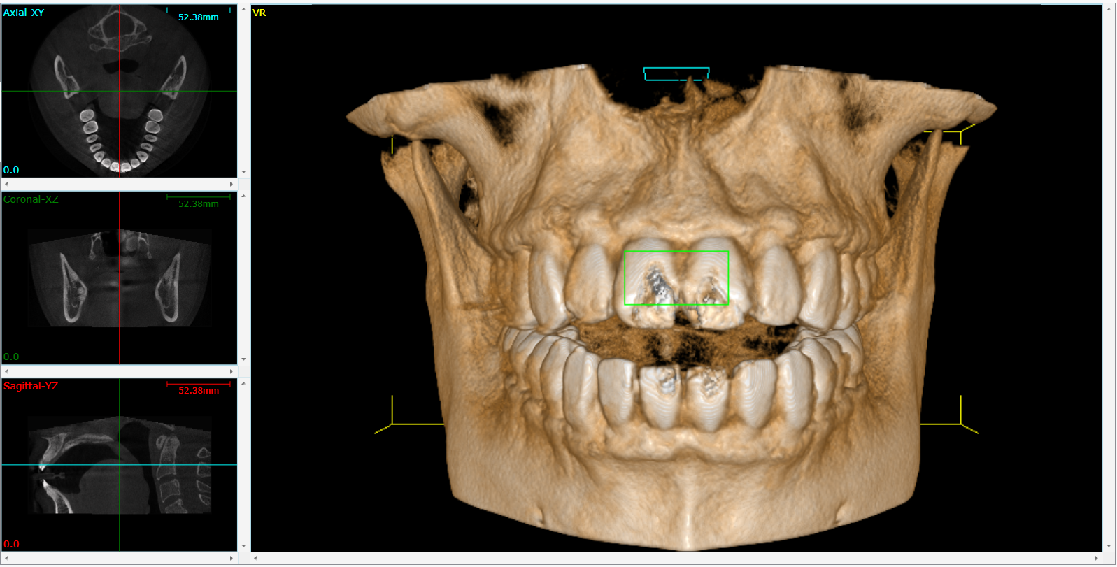
Exploring the image. A qualified dentist can then use the 3D model to see a number of details. For example, they can see different 2D or 3D views; they can zoom in or out; or they can get a full panoramic view of the mouth. This image is extremely helpful for planning surgery, constructing implants, or creating orthodontic appliances.
Interested in Finding Out More?
You can reach out to us with your questions about CBCT. Not in Florida? Try finding a dentist using our local dentist finder.